[논문/International Journal of Molecular Sciences] Highly Bright Silica-Coated InP/ZnS Quantum Dot-Embedded Silica Nanoparticles as Biocompatible Nanoprobes
작성자
관리자작성일자
2022-09-20 15:50조회수
54KIURI 김형모 참여연구원 논문
International Journal of Molecular Sciences. 2022 Sep 19; doi: https://doi.org/10.3390/ijms231810977
Highly Bright Silica-Coated InP/ZnS Quantum Dot-Embedded Silica Nanoparticles as Biocompatible Nanoprobes
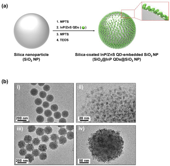
Quantum dots (QDs) have outstanding optical properties such as strong fluorescence, excellent photostability, broad absorption spectra, and narrow emission bands, which make them useful for bioimaging. However, cadmium (Cd)-based QDs, which have been widely studied, have potential toxicity problems. Cd-free QDs have also been studied, but their weak photoluminescence (PL) intensity makes their practical use in bioimaging challenging. In this study, Cd-free QD nanoprobes for bioimaging were fabricated by densely embedding multiple indium phosphide/zinc sulfide (InP/ZnS) QDs onto silica templates and coating them with a silica shell. The fabricated silica-coated InP/ZnS QD-embedded silica nanoparticles (SiO2@InP QDs@SiO2 NPs) exhibited hydrophilic properties because of the surface silica shell. The quantum yield (QY), maximum emission peak wavelength, and full-width half-maximum (FWHM) of the final fabricated SiO2@InP QDs@SiO2 NPs were 6.61%, 527.01 nm, and 44.62 nm, respectively. Moreover, the brightness of the particles could be easily controlled by adjusting the amount of InP/ZnS QDs in the SiO2@InP QDs@SiO2 NPs. When SiO2@InP QDs@SiO2 NPs were administered to tumor syngeneic mice, the fluorescence signal was prominently detected in the tumor because of the preferential distribution of the SiO2@InP QDs@SiO2 NPs, demonstrating their applicability in bioimaging with NPs. Thus, SiO2@InP QDs@SiO2 NPs have the potential to successfully replace Cd-based QDs as highly bright and biocompatible fluorescent nanoprobes.
Keywords: quantum dots (QDs); silica-coated InP/ZnS QD-embedded silica nanoparticles; biocompatible nanoprobes; photoluminescence (PL); syngeneic mice; in vivo; bioimaging
